


Sparks literally fly when a sperm and an egg hit it off. Chemists helped detect how the fertilized mammalian egg releases from its surface billions of zinc atoms in "zinc sparks," one wave after another.
The University of Texas at Austin's Emily Que, an assistant professor of chemistry, was lead author on a new study that describes the cutting-edge technology a research team used to become the first to capture images of these molecular fireworks and to pinpoint the origin of the zinc sparks: tiny zinc-rich packages just below the egg's surface. The beautiful display, captured on video, has garnered widespread attention online this week, with sites such as BuzzFeed and the Huffington Post weighing in on how sparks really do fly when egg and sperm hook up.
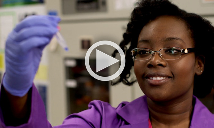
The video shows zinc being released from the egg during a zinc spark. Hot spots of fluorescence at the cell surface demonstrate how groups of zinc-rich packages are released from the egg during fertilization, resulting in a 'zinc spark.' Zinc fluctuations play a central role in regulating the biochemical processes that ensure a healthy egg-to-embryo transition. The new research should prove useful in improving in vitro fertilization outcomes. Because eggs that release more zinc would signal healthier embryos, the ability to identify those fertilized eggs early on would make it easier for doctors to know which embryos to use in fertility treatments.
The study, which took place at Northwestern University where Que was a post-doctoral researcher, also involved experts from the U.S. Department of Energy's Advanced Photon Source (APS). The research was published this week by the journal Nature Chemistry in a paper that describes how the researchers used sensitive imaging methods to see and count individual zinc atoms in egg cells and visualize zinc spark waves in three dimensions. Corresponding authors were obstetrics and gynecology expert Teresa Woodruff and chemist Thomas V. O'Halloran, both of Northwestern.
After inventing a novel vital fluorescent sensor for live-cell zinc tracking, scientists discovered that, at the point of fertilization, an egg releases thousands of packages that each dump roughly a million zinc atoms, followed by several more bursts of zinc release that spark like fireworks. The study establishes how eggs compartmentalize and distribute zinc to control the developmental processes that allow the egg to become a healthy embryo.
Zinc is part of a master switch that controls the decision to grow and change into a completely new genetic organism, making the new finding about how it works significant for medical purposes such as needing to identify healthy embryos in in vitro fertilization (IVF). Embryo weakness is one of the leading causes for implantation failure in IVF, a procedure that now results in implantation and later healthy delivery of a child only about 1 out of every 4 times. Because the procedure is both expensive and often heart-wrenching for would-be parents, improving IVF would be a crucial breakthrough for families struggling with fertility.
The National Institute of General Medical Sciences, the National Institute of Child Health and Human Development and the W.M. Keck Foundation supported the research described in the paper, "Quantitative mapping of zinc fluxes in the mammalian egg reveals the origin of fertilization-induced zinc sparks." Other authors besides Que, Woodruff and O'Halloran were engineer Vinayak P. Dravid and physicist Stefan Vogt, and Reiner Bleher, Francesca E. Duncan, Betty Y. Kong, Seth A. Garwin and Amanda R. Bayer, of Northwestern; and Sophie C. Gleber and Si Chen, of Argonne National Laboratory.
This post was adapted from a Northwestern University press release.
Using cutting-edge technology they developed, the team is the first to capture images of these molecular fireworks and pinpoint the origin of the zinc sparks: tiny zinc-rich packages just below the egg’s surface.
Zinc fluctuations play a central role in regulating the biochemical processes that ensure a healthy egg-to-embryo transition, and this new unprecedented quantitative information should be useful in improving in vitro fertilization methods.
“The amount of zinc released by an egg could be a great marker for identifying a high-quality fertilized egg, something we can’t do now,” said Teresa K. Woodruff, an expert in ovarian biology and one of two corresponding authors of the study. “If we can identify the best eggs, fewer embryos would need to be transferred during fertility treatments. Our findings will help move us toward this goal.”
Woodruff is a Thomas J. Watkins Memorial Professor in Obstetrics and Gynecology and director of the Women’s Health Research Institute at Northwestern University Feinberg School of Medicine.
The study is published today (Dec. 15) by the journal Nature Chemistry, provides the first quantitative physical measurements of zinc localization in single cells in a mammal.
The research team, including experts from the U.S. Department of Energy’s Advanced Photon Source (APS), developed a suite of four physical methods to determine how much zinc there is in an egg and where it is located at the time of fertilization and in the two hours just after. Sensitive imaging methods allowed the researchers to see and count individual zinc atoms in egg cells and visualize zinc spark waves in three dimensions.
After inventing a novel vital fluorescent sensor for live-cell zinc tracking, scientists discovered close to 8,000 compartments in the egg, each containing approximately one million zinc atoms. These packages release their zinc cargo simultaneously in a concerted process, akin to neurotransmitter release in the brain or insulin release in the pancreas.
These findings were further confirmed with chemical methods that trap cellular zinc stores and enable zinc mapping on the nanometer scale in a custom-designed electron microscope developed for this project with funding from the W.M. Keck Foundation. Additional high-energy X-ray imaging experiments at the APS synchrotron at Argonne National Laboratory enabled the scientists to precisely map the location of zinc atoms in two and three dimensions.
“On cue, at the time of fertilization, we see the egg release thousands of packages, each dumping a million zinc atoms, and then it’s quiet,” said Thomas V. O’Halloran, the other corresponding author. “Then there is another burst of zinc release. Each egg has four or five of these periodic sparks. It is beautiful to see, orchestrated much like a symphony. We knew zinc was released by the egg in huge amounts, but we had no idea how the egg did this.”
O’Halloran is a Charles E. and Emma H. Morrison Professor in Chemistry in the Weinberg College of Arts and Sciences and director of Northwestern’s Chemistry of Life Processes Institute.
The study establishes how eggs compartmentalize and distribute zinc to control the developmental processes that allow the egg to become a healthy embryo. Zinc is part of a master switch that controls the decision to grow and change into a completely new genetic organism.
The studies reported in Nature Chemistry are the culmination of six years of work and build on prior discoveries made by the Woodruff and O’Halloran labs using data from work performed at Northwestern and the APS. In previous studies in mouse eggs, this research team discovered the egg’s tremendous zinc requirement for reaching maturity. In addition, the researchers determined that an egg loses 10 billion of its 60 billion zinc atoms upon fertilization in a series of four or five waves called “zinc sparks.” Release of zinc sparks from the egg is essential for embryo formation in the two hours following fertilization.
“The egg first has to stockpile zinc and then must release some of the zinc to successfully navigate maturation, fertilization and the start of embryogenesis,” O’Halloran said. “But exactly how much zinc is involved in this remarkable process and where is it in the cell? We needed data to better understand the molecular mechanisms at work as an egg becomes a new organism.”
One major hurdle O’Halloran and Woodruff faced was the lack of sensitive methods for measuring zinc in single cells. To address this problem, they formed a collaborative team with other researchers in Northwestern’s Chemistry of Life Processes Institute to develop the tools they needed.
Key members of the team were Vinayak P. Dravid, the Abraham Harris Professor of Materials Science and Engineering at the McCormick School of Engineering and Applied Science, and Stefan Vogt, a physicist and group leader of microscopy at the Advanced Photon Source. Dravid and Vogt are co-authors of the paper.
“We had to develop a slew of methods to be convinced we were seeing the right thing,” O’Halloran said. “Science is about testing and retesting ideas. All of our complementary results point to the same conclusion: the zinc originates in packages called vesicles near the cell’s surface.”
The researchers currently are working to see if they can correlate zinc sparks with egg quality, information that would be key to improving fertility treatments.
Not only are these new imaging techniques important for describing the zinc spark, they can be applied to other cells that likely use zinc in a similar way, but whose workings remain elusive due to the lack of sensitive and specific tools. This study lays the groundwork for understanding how zinc fluxes can regulate events in multiple biological systems beyond the egg, including neurotransmission from zinc-enriched neurons in the brain and insulin-release in the pancreas.
The National Institutes of Health, Eunice Kennedy Shriver National Institute of Child Health and Human Development (grant U54HD076188) and the W.M. Keck Foundation supported the research.
The title of the paper is “Quantitative mapping of zinc fluxes in the mammalian egg reveals the origin of fertilization-induced zinc sparks.”
In addition to O’Halloran, Woodruff, Dravid and Vogt, other authors of the paper are lead author Emily L. Que, Reiner Bleher, Francesca E. Duncan, Betty Y. Kong, Seth A. Garwin and Amanda R. Bayer, of Northwestern; and Sophie C. Gleber and Si Chen, of Argonne National Laboratory.
- See more at: http://www.northwestern.edu/newscenter/stories/2014/12/stunning-zinc-fireworks-when-egg-meets-sperm.html#sthash.WMpG6wmU.dpufUsing cutting-edge technology they developed, the team is the first to capture images of these molecular fireworks and pinpoint the origin of the zinc sparks: tiny zinc-rich packages just below the egg’s surface.
Zinc fluctuations play a central role in regulating the biochemical processes that ensure a healthy egg-to-embryo transition, and this new unprecedented quantitative information should be useful in improving in vitro fertilization methods.
“The amount of zinc released by an egg could be a great marker for identifying a high-quality fertilized egg, something we can’t do now,” said Teresa K. Woodruff, an expert in ovarian biology and one of two corresponding authors of the study. “If we can identify the best eggs, fewer embryos would need to be transferred during fertility treatments. Our findings will help move us toward this goal.”
Woodruff is a Thomas J. Watkins Memorial Professor in Obstetrics and Gynecology and director of the Women’s Health Research Institute at Northwestern University Feinberg School of Medicine.
The study is published today (Dec. 15) by the journal Nature Chemistry, provides the first quantitative physical measurements of zinc localization in single cells in a mammal.
The research team, including experts from the U.S. Department of Energy’s Advanced Photon Source (APS), developed a suite of four physical methods to determine how much zinc there is in an egg and where it is located at the time of fertilization and in the two hours just after. Sensitive imaging methods allowed the researchers to see and count individual zinc atoms in egg cells and visualize zinc spark waves in three dimensions.
After inventing a novel vital fluorescent sensor for live-cell zinc tracking, scientists discovered close to 8,000 compartments in the egg, each containing approximately one million zinc atoms. These packages release their zinc cargo simultaneously in a concerted process, akin to neurotransmitter release in the brain or insulin release in the pancreas.
These findings were further confirmed with chemical methods that trap cellular zinc stores and enable zinc mapping on the nanometer scale in a custom-designed electron microscope developed for this project with funding from the W.M. Keck Foundation. Additional high-energy X-ray imaging experiments at the APS synchrotron at Argonne National Laboratory enabled the scientists to precisely map the location of zinc atoms in two and three dimensions.
“On cue, at the time of fertilization, we see the egg release thousands of packages, each dumping a million zinc atoms, and then it’s quiet,” said Thomas V. O’Halloran, the other corresponding author. “Then there is another burst of zinc release. Each egg has four or five of these periodic sparks. It is beautiful to see, orchestrated much like a symphony. We knew zinc was released by the egg in huge amounts, but we had no idea how the egg did this.”
O’Halloran is a Charles E. and Emma H. Morrison Professor in Chemistry in the Weinberg College of Arts and Sciences and director of Northwestern’s Chemistry of Life Processes Institute.
The study establishes how eggs compartmentalize and distribute zinc to control the developmental processes that allow the egg to become a healthy embryo. Zinc is part of a master switch that controls the decision to grow and change into a completely new genetic organism.
The studies reported in Nature Chemistry are the culmination of six years of work and build on prior discoveries made by the Woodruff and O’Halloran labs using data from work performed at Northwestern and the APS. In previous studies in mouse eggs, this research team discovered the egg’s tremendous zinc requirement for reaching maturity. In addition, the researchers determined that an egg loses 10 billion of its 60 billion zinc atoms upon fertilization in a series of four or five waves called “zinc sparks.” Release of zinc sparks from the egg is essential for embryo formation in the two hours following fertilization.
“The egg first has to stockpile zinc and then must release some of the zinc to successfully navigate maturation, fertilization and the start of embryogenesis,” O’Halloran said. “But exactly how much zinc is involved in this remarkable process and where is it in the cell? We needed data to better understand the molecular mechanisms at work as an egg becomes a new organism.”
One major hurdle O’Halloran and Woodruff faced was the lack of sensitive methods for measuring zinc in single cells. To address this problem, they formed a collaborative team with other researchers in Northwestern’s Chemistry of Life Processes Institute to develop the tools they needed.
Key members of the team were Vinayak P. Dravid, the Abraham Harris Professor of Materials Science and Engineering at the McCormick School of Engineering and Applied Science, and Stefan Vogt, a physicist and group leader of microscopy at the Advanced Photon Source. Dravid and Vogt are co-authors of the paper.
“We had to develop a slew of methods to be convinced we were seeing the right thing,” O’Halloran said. “Science is about testing and retesting ideas. All of our complementary results point to the same conclusion: the zinc originates in packages called vesicles near the cell’s surface.”
The researchers currently are working to see if they can correlate zinc sparks with egg quality, information that would be key to improving fertility treatments.
Not only are these new imaging techniques important for describing the zinc spark, they can be applied to other cells that likely use zinc in a similar way, but whose workings remain elusive due to the lack of sensitive and specific tools. This study lays the groundwork for understanding how zinc fluxes can regulate events in multiple biological systems beyond the egg, including neurotransmission from zinc-enriched neurons in the brain and insulin-release in the pancreas.
The National Institutes of Health, Eunice Kennedy Shriver National Institute of Child Health and Human Development (grant U54HD076188) and the W.M. Keck Foundation supported the research.
The title of the paper is “Quantitative mapping of zinc fluxes in the mammalian egg reveals the origin of fertilization-induced zinc sparks.”
In addition to O’Halloran, Woodruff, Dravid and Vogt, other authors of the paper are lead author Emily L. Que, Reiner Bleher, Francesca E. Duncan, Betty Y. Kong, Seth A. Garwin and Amanda R. Bayer, of Northwestern; and Sophie C. Gleber and Si Chen, of Argonne National Laboratory.
- See more at: http://www.northwestern.edu/newscenter/stories/2014/12/stunning-zinc-fireworks-when-egg-meets-sperm.html#sthash.WMpG6wmU.dpuf


Comments 2
Well written article documenting a potentially very 'lifechanging' research result.
for the untrained eye it tool several reruns of the video clip to understand and see what was being filmed; slowing the video down would have helped
Exciting research, especially for those of us that work in reproductive medicine.
Just a quick comment regarding the purported IVF success rate of "1 in 4 times." That is is a seriously outdated statistic.
With modern IVF, we test embryos for their chromosomal constitution prior to embryo transfer. The overall implantation rate of a single chromosomally normal embryo is between 40-80%, depending on the age of the egg provider. Lower miscarriage rates and no twins as well. Research like this may help to determine if zinc release (cortical granule reaction?) has anything to do with observed differences in IVF success due to female age in the setting of otherwise proven chromosomally norma embryos.