


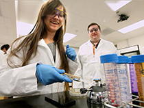


Neurobiologist Kristen Harris recently joined the Center for Learning and Memory (CLM), and she brings with her a pioneering technique to study how neurons change and how the changes relate to learning and memory.
Harris has a special interest in the structure and function of protrusions on neurons called dendritic spines. The spines receive chemical signals passing from the axon of one neuron to the dendrites of another. These places of connection between neurons, called synapses, change as the brain learns and remembers.
Some of Harris’s research involves studying images of dendrites to learn how the spines grow and diminish in response to stimuli. She looks at the brains of rats of varied ages to understand how long-term potentiation, a cellular model of learning, changes as organisms mature.
One of the most important discoveries from the Harris lab is that rapid changes in the structure of synapses are accompanied by local changes in the ability of dendrites to manufacture new proteins. These changes occur within minutes of being stimulated, much more rapidly than previously held theories of several hours.
To see these changes, Harris slices portions of the rat brain, usually from the hippocampus, into very thin sections–about 40 nanometers thick—and creates images of them with an electron microscope.
She and her colleagues then meticulously measure and outline the various parts of neurons cross-sectioned in each slice. Some of the structures are as small as 10 to 20 nanometers. Finally, with the help of a computer, the images are stacked atop one another like cards in a deck to create a complete 3D reconstruction of specific sets of synapses, dendrites and axons.
“It’s like piecing a puzzle back together,” says Harris. “Only you can get lost in this puzzle and it takes both biological knowledge about how the structures relate to one another, and patience to understand and reconstruct what one is looking at.”
Before arriving at the CLM in Fall 2006, Harris directed laboratories at the Medical College of Georgia, Boston University, and Harvard Medical School. She was drawn to the CLM, Department of Neurobiology and Institute for Neuroscience by the chance to collaborate with the university’s cutting-edge faculty and by the opportunity to do research with the next generation of neuroscientists.
“I want to interact with the larger student population,” Harris says, “and to help advance a terrific degree program in neuroscience.”
Learn more at: synapse-web.org.
Written by Patrick Brendel.


Comments